Secondary Ion Mass Spectrometry Instrumentation TutorialSIMS HistoryBombardment of a sample surface with a primary ion beam followed by mass spectrometry of the emitted secondary ions constitutes secondary ion mass spectrometry (SIMS). The first inklings of the SIMS process came when early mass spectroscopists noticed that ions from instrument construction materials were produced by ion sources. Later experiments extracted ions from the sources and accelerated them onto the sample, thereby producing the first SIMS primary ion beam. The first SIMS instrument was constructed under a NASA contract in the early 1960's to analyze moon rocks. When it performed better than expected, exact copies of the prototype were introduced into the market place. The use of SIMS for materials characterization has grown steadily during the intervening 30 years. SIMS measures trace levels of all elements in the periodic table. SIMS also provides lateral and depth distributions (microanalysis) of these elements within a sample. The electronic materials industries (semiconductors, optoelectric devices, etc.) are the largest users of SIMS. The geological community also uses SIMS for laterally resolved isotopic and elemental measurements.
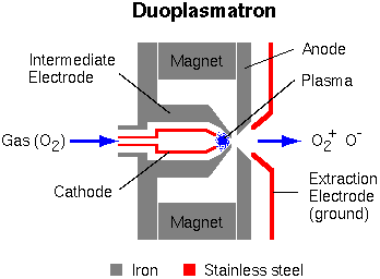
SIMS Primary Ion SourcesTypical SIMS instruments use either a duoplasmatron or a surface ionization primary ion source (or both). The duoplasmatron can operate with virtually any gas, but oxygen is the most common because oxygen implantation into the sample surface enhances ionization efficiency for electropositive elements. Before this oxygen enhancement effect was discovered, argon was commonly used. The oxygen plasma within the duoplasmatron source contains both O- and O2+, and either can be extracted.
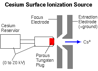
The cesium surface ionization source produces Cs+ ions as Cs atoms vaporize through a porous tungsten plug.
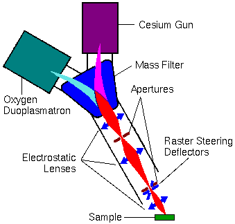
SIMS Primary Ion ColumnPrimary ions are extracted from the sources and passed to the sample through the primary ion column. The column usually contains a primary beam mass filter that transmits only ions with a specified mass-to-charge (m/z) ratio. This mass filter eliminates impurity species in the beam. For example, Cr, Fe, and Ni ions sputter from stainless steel surfaces within a duoplasmatron. Without a primary beam mass filter, these metal contaminants deposit onto the sample surface, raising the detection limits for stainless steel elements.
In the figure above, the electromagnetically active components are shown in blue. The ion beam trajectories (indicated in red) are greatly exaggerated in the lateral directions. The electrostatic lenses and the apertures control the intensity and width of the primary ion beam. Several aperture diameters are usually available at each aperture location. The primary beam intensity can be reduced by defocusing the ion beam onto the back of the first aperture (nearest the magnet). A narrow beam (at the sample) results from defocusing the ion beam (with the middle lens) onto the back of the second aperture, and then adjusting the last lens to transfer the image of the cross-over from behind the aperture onto the sample. Electrostatic deflectors steer the primary beam in a raster pattern onto the sample. A finely focused and rastered primary ion beam delivers uniform primary beam intensity to an area on the sample. This leads to flat bottom sputter craters. The best depth resolution in a depth profile results when the secondary ions are sampled from the flat bottom of such a crater without contributions from the crater edges. Other deflectors (not shown) are located near the apertures. They help tune the primary beam through the middle of the electrostatic lenses.
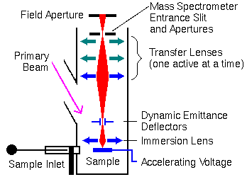
Secondary Ion Extraction and TransferSecondary ions are extracted from the sample as they are produced. If large mass spectrometer components are held at ground potential, the sample must be held at high voltage, the accelerating potential. The secondary ions accelerate toward the ground plate of an electrostatic lens. This first lens is called the immersion or ion extraction lens. The second (transfer lens) focuses the ion beam onto the mass spectrometer entrance slits or aperture. This two lens system constitutes an ion microscope. The secondary ions could be projected onto an image detector for viewing the sample surface. Different transfer lenses produce different magnifications.
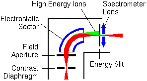
In the figure above, the electromagnetically active components are shown in blue. The ion beam trajectories (indicated in red) are greatly exaggerated in the lateral directions. The field aperture is located approximately at the point where the ion beam image comes into focus. The entrance aperture is sometimes called a contrast diaphragm. Smaller aperture diameters intercept ions with off-axis energy components. This reduces image aberrations but unfortunately it also reduces secondary ion intensity. Ions that arise from off the secondary ion optical axis contribute to lower mass resolution. These off-axis ions arise because the primary beam raster pattern sputters an area rather than the single point where the axis intercepts the sample. The dynamic emittance deflectors adjust the secondary ion beam back on-axis. The deflectors operate in synchrony with the primary beam raster generator to provide continuous adjustment. Ion Energy AnalyzersElectrostatic energy analyzers bend lower energy ions more strongly than higher energy ions. The sputtering process produces a range of ion energies. An energy slit can be set to intercept the high energy ions (shown in green).
In the figure above, the electromagnetically active components are shown in blue. The ion beam trajectories (indicated in red) are greatly exaggerated in the lateral directions. Voltage offset is a strategy for enhancing monatomic ions over multiatomic. The monoatomic ions have higher energy distributions. If the accelerating voltage is lowered (offset), more of the atomic ions still have enough energy to pass through the energy slits. In a typical SIMS experiment, the accelerating voltage is 4.5 kV, and the offset is 50 V. The inner jaw of the slits intercepts most (low energy) multiatomic ions. Both monatomic and multiatomic ion intensities are reduced in a voltage offset measurement, but multiatomic ions relatively more than monoatomic. The inner and outer sector electrodes have voltages of opposite polarity. Their magnitude is about 10% of the ion accelerating voltage. The ion image comes into focus, producing a virtual image inside the electrostatic sector behind the field aperture. The active surfaces of the electrostatic sector are spherical. This geometry transfers the image to the mass analyzer with minimal distortion. The spectrometer lens adjusts the ion beam focus (cross over) to meet the input requirements of the mass analyzer.
Mass AnalyzersDynamic SIMS instruments use two kinds of mass analyzers, magnetic sector and quadrupole. Magnetic sector instruments are most common. As the ion beam passes through the magnetic field, the particles are acted on by a force at right angles, both to the direction of motion and to the direction of the magnetic field. The following equation shows the relationship between the magnetic field (B), the ion accerating voltage (V), the mass-to-charge ratio (m/q), and the radius of ion curvature (r) in the magnetic field. In atomic units, m/q becomes m/z where z is the number of charges on the ion.
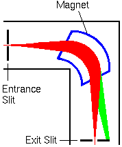
Magnetic sector mass analyzer is shown in blue. The ion beam trajectories (indicated in red) are greatly exaggerated in the lateral directions. Modern mass spectrometers use non-normal pole faces for entrance and exit of the ion beam to the magnetic sector. The fringings fields in this configuration compress the ion beam in the vertical direction (in and out of the screen) as it passes through the sector. Fewer ions strike metal surfaces and the ion beam focuses better at the exit slit with non-normal pole faces. The entrance and exit slits can be arranged at ion beam crossovers for the cleanest separation (highest mass resolution) between ions with similar m/z values. The green part of the beam represents ions with higher m/z values that do not pass through the spectrometer.
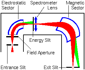
The combination of a magnetic and an electrostatic sector produces a double focusing instrument. A magnetic analyzer, by itself, introduces chromatic aberrations into an ion beam with dispersed ion energies. These aberrations reduce mass resolution. In a series arrangement of one electrostatic and one magnetic sector, the energy dispersion of the electrostatic sector can just compensate the energy dispersion of the magnet. The system will have the mass dispersive properties of the magnet, except that it will produce higher mass resolution. The spectrometer lens adjusts the cross-over from the electrostatic sector to the location required for the magnetic sector.
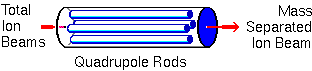
Quadrupole mass analyzers were invented in 1953. Many kinds of analysis, including SIMS, employ quadrupoles. Ideally, the rods have hyperbolic shapes, but this geometry can be approximated with closely spaced circular rods. In a typical quadrupole spectrometer, the rods are 1 cm in diameter and 20 cm long. In the diagram, ions enter from the left at a relatively low energy (~25 eV). Since SIMS ions can have a wider energy range than 25 eV, electrostatic sectors usually precede the quadrupole.
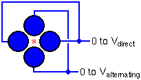
Alternating and direct voltages on the rods cause the ions to oscillate after entering the quadrupole. For a given set of voltages, Ions with a single mass-to-charge ratio undergo stable oscillation and traverse through the rods. All other ions have unstable oscillations and strike the rods. The alternating frequency and the ratio between the alternating and direct voltages remain constant. Scanning the voltages scans the mass spectrum.
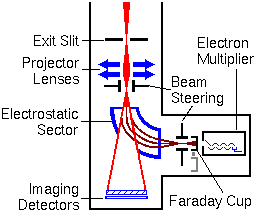
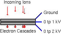
Secondary Ion DetectorsThe most widely used SIMS instruments have as many as four detectors. These include an ion counting electron multiplier, a Faraday cup, and two ion image detectors. The following figure shows the arrangement of detectors. The ion counting electron multipliers are the most sensitive detectors. They must be protected from intense ion beams. The Faraday cup detector moves on a solenoid to cover the electron multiplier when the incoming ion signal is too high. High energy neutral species form by charge exchange when an ion beam strikes a surface. These neutrals contribute noise to the ion signal. If an electrostatic sector precedes the electron multiplier, the neutrals can be eliminated from the ion signal. Quadrupole mass analyzers also use electrostatic sectors or deflectors to minimize the contributions of high energy neutral species to the ion signal. The ion beam passes through a small hole in the electrostatic sector when the sector is deactivated. This path leads to dual microchannel plate and resistive anode encoder image detectors. The projector lenses bring an image of the sample into focus on the image detectors.
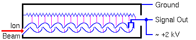
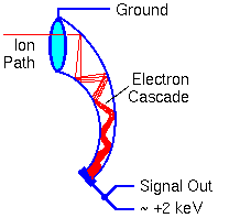
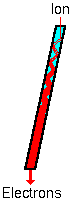
Electromagnetically active components are shown in blue. The ion beam trajectories (indicated in red) are greatly exaggerated in the lateral directions. In particular, the image detectors are smaller and the path through the electrostatic analyzer is narrower. The ions pass through a much smaller hole in the sector. Electron MultipliersAn electron multiplier consists of a series of electrodes called dynodes, each connected along a resistor string. The signal output end of the resistor string attaches to positive high voltage. The other end of the string goes to the electron multiplier case and ground.
The dynode potentials differ in equal steps along the chain. When a particle (electron, ion, high energy neutral, or high energy photon) strikes the first dynode it produces secondary electrons. The secondary electrons are accelerated into the next dynode where each electron produces more secondary electrons. A cascade of secondary electrons ensues. The dynode acceleration potential controls the electron gain.
Electron multipliers can also be made from continuous dynode materials rather than discrete dynodes. This glassy material contains lead that provides conductivity comparable to the resistor chain in the discrete dynode electron multipliers. Most SIMS measurements use electron multipliers operating with sufficiently high gain to produce a detectable pulse for every ion arrival. Pulse counting is the most sensitive ion detection method. Detector noise arises from stray ions and cosmic rays, but these signals are normally less than one count per second. In order to detect both positive and negative ions, the electron multiplier case stands at ground potential. The output end of the resistor chain must operate at high positive potential. This requires that the output pulse be capacitively coupled to the detector electronics. The detector electronics require a recovery time (dead time) after an ion arrival before a second ion can be detected. The detector dead time limits the measureable ion arrival rate to around 1e6 counts per second. Thus the electron multiplier dynamic range extends from below 1 to about 1e6 ion counts per second. Pulse counting detectors follow Poisson statistics which require that each ion arrives independently of all other ions. A measurement consists of counting ions for a fixed period of time and the result takes the form of a counting number, n. The standard deviation of the measurement is equal to the square root of the number of counts. The second equation shows the standard deviation relative to the signal.
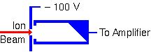
Faraday Cups
A Faraday cup is just an electrode from which electrical current is measured while a charge particle beam (electrons or ions) impinges on it. The shape helps minimize loss of secondary electrons that would alter the current measurement. A deep cup with an electron repeller plate minimizes secondary electron loss.
Ion Image Detectors
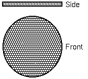
Ion image detectors depend on microchannel plate electron multiplier arrays. These plates consist of large arrays of small channel electron multipliers. SIMS instruments typically use round arrays with about 2000 channels across a diameter. Each channel is 10 microns in diameter. Channels are located on 12 micron centers and the total array is 25 mm in diameter.
Each channel has dimensions of 10 x 400 micron. The channels are 7 degrees from perpendicular to the array surface. The voltage across single channel plate can produce gains has high as 1e5.
For SIMS use, two microchannel plates combine to easily produce gains of 1e6. Two kinds of anode provide either direct visualization, or computer compatible ion position data. Two microchannel plates followed by a phosphor screen for visuallizing the electron cascade provides an easy way to monitor the secondary ion beam. SIMS analysts call this combination a dual microchannel plate (DMCP). As the electrons are accelerated into the phosphor anode, they generate more than one photon per electron. Thus, the anode provides additional gain.
|